
Live Cell Evaluation of Cell Cycle Inhibitors - ImageXpress® Micro
Foreword
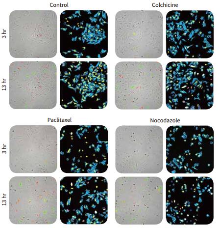
Monitoring the effects on cell cycle changes plays an important role in tumor development and drug discovery. For example, compounds known to inhibit mitosis are mostly used to slow the growth of tumor cells. Screening for high content of living cells has been validated to distinguish each cycle of cells. This technology, in combination with the BacMam transfection system, allows cells to express two cell cycle-associated fluorescent fusion proteins.
The delayed monitoring experiment lasted 2-3 days and the cells were placed in the environmental control chamber of the ImageXpress ® Micro high-content imaging and analysis system. The entire experiment maintained the normal growth environment of living cells. Live cells and fluorescent live cell images are acquired simultaneously at pre-set intervals. This complete protocol consists of a pre-built module that analyzes time-lapse images, identifies all cells based on brightfield images, and differentiates between cell cycles based on fluorescent protein expression.
Visualizing the fluorescent protein indicates the cell's cycle
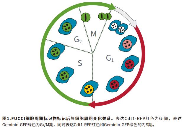
Using real-time monitoring of cell cycle Premo TM FUCCI cell cycle indicator (Thermo Fisher). Prior to seeding, cells were transfected with fluorescently labeled cell cycle associated proteins, geminin and Cdt1. Geminin is green GFP, Cdt1 is red RFP, and the shooting channels are FITC and TRITC. Since both geminin and Cdt1 are expressed only in specific cycles, their expression in the nucleus can indicate the cell cycle in which the cell is located (Fig. 1).
Advantage
- Live cells continue to grow in the instrument for up to 72 hours
- Reduce experimental operation time
- Identify cells without labeling in bright field
- Detection of cyclical changes using cell cycle transfection reagents
method
1. Prepare the cell suspension, mix 40,000 cells/mL Hela cell suspension with 30 granule/cell concentration of FUCCI reagent, seed in 96-well plate at a concentration of 4,000 cells/well, and place the plate at 37 °C, 5 Adhere for 8 hours in the environment of %CO2.
2. Treat cells with different concentrations of mitotic inhibitor and place the plates into the ImageXpressMicro system.
3. The system automatically takes a picture every 2-3 hours, using a 20x PlanApo objective, and simultaneously acquires the bright field and two fluorescent channels FITC and TRITC images. The experiment lasted 48-72 hours and the cells could complete 1-2 divisions.
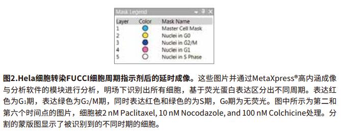
4. Analyze delayed experimental images using MetaXpress® high-content imaging and analysis software for user-defined modules.
Bright field recognition cells, fluorescent proteins distinguish cell cycle
Living cells use the FUCCI cell cycle indicator, and changes in cell cycle after compound treatment can be monitored in real time, and more information can be obtained than the endpoint method for detecting fixed cells. FUCCI transfected Hela cells in cell cycle indicator cell high-content in real-time image photographed, and analyzed by MetaXpress ® high content imaging and analysis software modules, bright field identify all cells, expressing a fluorescent protein-based distinction Different periods. The expression of red is G1 phase, the expression of green is G 2/M phase, and the expression of red and green is S phase (Fig. 2). Cells in the G0 phase have no fluorescent signal, but are recognized in the bright field and counted in the total number of cells. In addition, dividing cells can also be recorded.
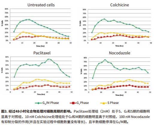
Cell cycle inhibitors can cause cells to stagnate in specific cycles
The analysis process accelerates the analysis speed using the MetaXpress PowerCore parallel processing software. After 24 hours of compound treatment, most of the cells stayed in the G0 phase, and only a few cells expressed the cell cycle marker protein. Cells in paclitaxel and nocodazole have certain cells arrested in the G2/M phase (Fig. 3).
to sum up
Premo TM FUCCI binding configuration environment indicator cell cycle control ImageXpressMicro MetaXpress imaging system and software, enables efficient cycle accurate measurement of viable cells. High-throughput screening technology provides scientists with a fast, automated method for quantitatively analyzing cell cycles based on images and is able to fully record cell cycle changes over time. In addition, MetaXpress software recognizes cells in brightfield images, avoiding the toxicity of dyes to living cells. A statistically significant quantitative analysis of the same standard can be used to evaluate parameters of the effects of multiple compounds on cell cycle at different concentrations.

Walnut Kernels,Walnut Kernel In Halves,Walnuts In Shell,Walnut Kernels Light Pieces
Weishan Yuanxing Walnuts Co.,Ltd. , https://www.walnutsxxy.com