introduction
Many types of nanoparticles can be used as delivery vehicles, and specific receptors can be designed and constructed to increase payloads by operating in a "hidden mode" and to extend the potency, reduce side effects, increase intake and Efficacy and so on.
Structures such as liposomes have been attracting researchers for many years. The use and potential of liposomes (Figure 1) as a drug delivery system is becoming increasingly prominent. The reason for this is obvious:
• Prevents the action of metabolic enzymes by liposome administration
• Increase the solubility of fat-soluble drugs
• Targeting drugs to specific areas by adding ligands
• Liposomes are easily absorbed by cells
• Drug release rate can be controlled by liposome selection
• The use of liposomes as a vehicle for drug delivery may reduce the amount of drug used, reduce toxicity and reduce side effects. Moreover, gene therapy drugs are also likely to be delivered through liposomes.
It has been gradually discovered that the size of liposomes is an important factor affecting the therapeutic efficacy. The size of the liposomes used for administration may affect the time it circulates and stays in the blood, the targeted efficacy, the rate of absorption (endocytosis) of the cells, and ultimately the successful release of its payload. Particle size is also extremely important for nanoscale polymer capsule delivery systems. Therefore, accurate measurement monitoring of its size is essential.
Figure 1: Representative structure of liposomes
Determination of liposome size using NanoSight NTA
The NanoSight instrument accurately and quickly measures the size of liposomes in water, requiring only a small sample to prepare samples. The system enables visualization of individual liposomes in suspension and tracks its Brownian motion. Based on the individual particles, the particle size distribution is known in a matter of seconds.
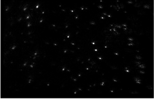
Figure 2: Typical image of liposomes given by the NanoSight instrument.
The NanoSight observation unit provides a unique viewing field for nanoparticles (Figure 2), while the Nanoparticle Tracking and Analysis Software Kit (NTA2.0) can be used to accurately determine the size of liposomes (Figure 3).
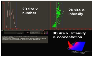
Figure 3: NTA results page generated by bimodal nanoparticle samples
Synchronous multi-parameter real-time analysis of nanoparticles
Nanoparticle refractive index
Thanks to the unique NanoSight technology that enables simultaneous and separate visualization of nanoparticles, more information about any given nanoparticle can be obtained.
The NanoSight system is capable of detecting the true particle size distribution of a sample based on quantity and a series of statistical measurements.
It also measures the relative light scattering intensity of the particles and plots the data against the independently obtained particle size measurements. This allows for more careful resolution of particles of different refractive indices or material compositions based on the superior particle size resolution provided by the NTA.
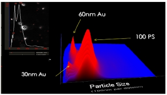
The following example shows a 100 nm polymer nanoparticle mixture with a relatively low refractive index (and therefore poor scattering) that can be easily distinguished from a mixture of seemingly smaller 30 nm and 60 nm metal nanoparticles.
Figure 4: NTA results showing 3 peaks: 30nm gold, 60nm gold and 100nm polystyrene
Please note that the 60nm peak is more susceptible to scattering. The vertical axis in these figures is the particle concentration and the quantity frequency distribution can be obtained directly from the sample.
This unique property makes it possible for the user to study whether nanoscale drug delivery structures such as liposomes will change in their contents. For example, the empty liposome's Ri value (light scattering ability) may be lower than loading. Liposomes of higher Ri value materials. This will allow them to be resolved even if the sizes are very similar.
Nanoparticle fluorescence
The replacement of a standard 635 nm red laser with different laser wavelengths (405 nm blue and 532 nm green laser) can stimulate a suitable fluorophore that is attached to or contained within a liposome or similar delivery vehicle.
Figure 5
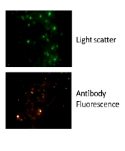
In the following examples, microvesicles and nanovesicles have been labeled with a suitable fluorophore using surface-specific antibodies, thus distinguishing specific subpopulations of identified nanovesicle particles from unlabeled.
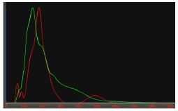
A similarly sized peak as shown in the figure below indicates that almost all of the vesicles seen in the light scattering mode (green line) have been labeled with a fluorophore (red line, as visualized at the appropriate excitation wavelength).
Figure 6: NTA results in light scattering mode (green line) and fluorescence mode (red line).
Sarms
Nonsteroidal SARMS are alternatives to steroids, also known as selective estrogen receptor modulators. Unlike steroids, SARMS stimulates muscle more directly and more strongly than steroids. The stronger the targeting, the more targeted. SARMS is an optimized version of steroid substances that have steroid-like effects in the treatment of certain diseases. It may completely replace steroids in the future as a new and more effective drug.
Our company specializes in providing sarms series products
MK677
MK2866
GW501516
YK11
SR9009
RAD140
S4
S23
LGD4033
GW0742
We can provide liquid, powder, capsule, etc. Welcome to inquiry

MK677
MK2866
GW501516
YK11
SR9009
RAD140
S4
S23
LGD4033
GW0742 powder

MK677
MK2866
GW501516
YK11
SR9009
RAD140
S4
S23
LGD4033
GW0742 capsules

Mk 677 Tablets,Mk 677 Pills,Sarms Liquid And Powder,Muscle Building Powder Swarm
XI AN RHINE BIOLOGICAL TECHNOLOGY CO.,LTD , https://www.rhinebiotech.com